


 The coxal (hip) joint is a synovial ball-and-socket joint formed by the rounded head of the femur (thigh bone) connecting into a cavity in the hip bone called the acetabulum. The left and right hip joints are located laterally on each side of the pelvis.
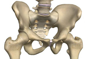
The two coxal (hip) bones fuse together to the lower front at the pubic symphysis, forming the pelvic girdle. To the upper rear, they connect with the sacrum, at the sacroiliac joint. The entire structure of hips, sacrum and pubic symphysis is known as the bony pelvis. The pelvis is a key part of the human skeleton, as it provides a strong support structure on which the spinal column can sit, while at the same time articulating with the legs, linking the lower limbs with the axial skeleton.
Each hip bone is itself comprised of three bones – the ilium, ischium and pubis. At birth these are separated by cartilage but they later fuse together to function as a single bone. The ilium is the upper part of the hip bone that connects with the sacrum at the sacroiliac joint. The ischium is located underneath the ilium and to the rear, whereas the pubis forms the lower front section. Both the pubis and ischium create the obturator foramen, which are the characteristic large holes which can be seen to either side of the lower part of the pelvis. All three bones form part of the acetabulum.
The hip joint allows for a range of movement of the leg including flexion (raising up), extension (straightening out), abduction (movement of the leg sideways away from the body), adduction (sideways movement back towards the body), external (lateral) rotation (circular movement away from the body) and internal (medial) rotation (circular movement towards the body). Around the joint, strength and support is provided by the fibers of the articular capsule and five sets of ligaments (the iliofemoral, pubofemoral, ischiofemoral, transverse ligament of the acetabulum and femur head ligament). The articular capsule extends from the edge of the acetabulum to the neck of the femur and is one of the strongest structures in the human body. This, and the fact that the rim of the acetabulum is narrower than the head of the femur, means that dislocation of the hip occurs very rarely.
There are a number of gender-based differences in the structure of the hip bone. These largely result from adaptations of the female pelvis for pregnancy and childbirth. The female pelvis is wider and shallower than that of the male. This provides for a larger pelvic space to permit the passage of the head of a full-term infant. In addition to being narrower than the female pelvis, the male hip bones tend to be larger, heavier and have greater surface markings.